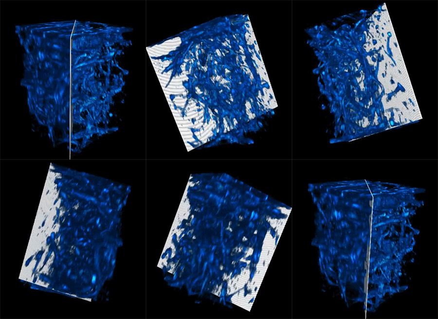
Metabolic imaging is a noninvasive technique that allows medical professionals and researchers to examine living cells utilizing laser light, which can assist them examine illness development and therapy actions.
Yet light scatters when it beams right into organic cells, restricting exactly how deep it can permeate and interfering with the resolution of caught photos.
Currently, MIT scientists have actually established a brand-new strategy that greater than increases the typical deepness limitation of metabolic imaging. Their technique likewise improves imaging rates, generating richer and much more thorough photos.
This brand-new strategy does not need cells to be preprocessed, such as by sufficing or discoloring it with dyes. Rather, a specialized laser lights up deep right into the cells, creating particular inherent particles within the cells and cells to send out light. This removes the demand to modify the cells, supplying a much more all-natural and exact depiction of its framework and feature.
The scientists attained this by adaptively tailoring the laser light for deep cells. Utilizing a lately established fiber shaper– a tool they manage by flexing it– they can tune the shade and pulses of light to reduce spreading and take full advantage of the signal as the light journeys deeper right into the cells. This enables them to see a lot even more right into living cells and capture more clear photos.
 appears today in Science Advances” This job reveals a substantial renovation in regards to deepness infiltration for label-free metabolic imaging. It opens up brand-new methods for researching and discovering metabolic characteristics deep in living biosystems,” claims Sixian You, assistant teacher in the Division of Electric Design and Computer Technology( EECS ), a participant of the Lab for Electronic devices, and elderly writer of a paper on this imaging strategy.
She is signed up with on the paper by lead writer Kunzan Liu, an EECS college student; Tong Qiu, an MIT postdoc; Honghao Cao, an EECS college student; Follower Wang, teacher of mind and cognitive scientific researches; Roger Kamm, the Cecil and Ida Eco-friendly Distinguished Teacher of Biological and Mechanical Design; Linda Griffith, the College of Design Teacher of Training Development in the Division of Biological Design; and various other MIT associates. The research study
.
Laser-focused
This brand-new technique drops in the classification of label-free imaging, which implies cells is not discolored ahead of time. Tarnishing produces comparison that assists a professional biologist see cell cores and healthy proteins much better. Yet discoloring commonly needs the biologist to area and cut the example, a procedure that frequently eliminates the cells and makes it difficult to examine vibrant procedures in living cells.
In label-free imaging methods, scientists utilize lasers to light up certain particles within cells, creating them to send out light of various shades that expose numerous molecular components and mobile frameworks. Nonetheless, producing the perfect laser light with particular wavelengths and premium pulses for deep-tissue imaging has actually been testing.prior work The scientists established a brand-new method to conquer this constraint. They utilize a multimode fiber, a sort of fiber optics which can lug a substantial quantity of power, and pair it with a small tool called a “fiber shaper.” This shaper enables them to exactly regulate the light proliferation by adaptively transforming the form of the fiber. Flexing the fiber alters the shade and strength of the laser.
Structure on
, the scientists adjusted the very first variation of the fiber shaper for much deeper multimodal metabolic imaging.
” We intend to carry all this power right into the shades we require with the pulse buildings we need. This offers us greater generation performance and a more clear photo, also deep within cells,” claims Cao.
Once they had actually developed the controlled device, they established an imaging system to utilize the effective laser resource to create longer wavelengths of light, which are important for much deeper infiltration right into organic cells.
” Our team believe this modern technology has the prospective to dramatically progress organic research study. By making it budget-friendly and obtainable to biology laboratories, we want to equip researchers with an effective device for exploration,” Liu claims.
Dynamic applications
When the scientists evaluated their imaging tool, the light had the ability to permeate greater than 700 micrometers right into an organic example, whereas the very best previous methods can just get to around 200 micrometers.
” With this brand-new kind of deep imaging, we intend to take a look at organic examples and see something we have actually never ever seen prior to,” Liu includes.
The deep imaging strategy allowed them to see cells at several degrees within a living system, which can assist scientists examine metabolic modifications that take place at various midsts. On top of that, the quicker imaging rate enables them to collect even more thorough details on exactly how a cell’s metabolic rate impacts the rate and instructions of its activities.
This brand-new imaging technique can provide an increase to the research of organoids, which are crafted cells that can expand to simulate the framework and feature of body organs. Scientists in the Kamm and Griffith laboratories leader the advancement of mind and endometrial organoids that can expand like body organs for illness and therapy evaluation.
Nonetheless, it has actually been testing to exactly observe interior advancements without reducing or discoloring the cells, which eliminates the example.
This brand-new imaging strategy enables scientists to noninvasively keep an eye on the metabolic states inside a living organoid while it remains to expand.
With these and various other biomedical applications in mind, the scientists prepare to go for also higher-resolution photos. At the exact same time, they are functioning to produce low-noise laser resources, which can allow much deeper imaging with much less light dose.
They are likewise establishing formulas that respond to the photos to rebuild the complete 3D frameworks of organic examples in high resolution.
In the future, they want to use this strategy in the real life to assist biologists keep an eye on medicine action in real-time to assist in the advancement of brand-new medications.
” By making it possible for multimodal metabolic imaging that gets to much deeper right into cells, we’re supplying researchers with an unmatched capacity to observe nontransparent organic systems in their all-natural state. We’re thrilled to work together with medical professionals, biologists, and bioengineers to press the borders of this modern technology and transform these understandings right into real-world clinical innovations,” You claims.
” This job is amazing since it utilizes cutting-edge responses approaches to photo cell metabolic rate deeper in cells contrasted to present methods. These innovations likewise supply quick imaging rates, which was utilized to discover one-of-a-kind metabolic characteristics of immune cell mobility within capillary. I anticipate that these imaging devices will certainly contribute for finding web links in between cell feature and metabolic rate within vibrant living systems,” claims Melissa Skala, a detective at the Morgridge Institute for Study that was not entailed with this job.
” Having the ability to get high resolution multi-photon photos relying upon NAD( P) H autofluorescence comparison quicker and deeper right into cells unlocks to the research of a vast array of vital issues,” includes Irene Georgakoudi, a teacher of biomedical design at Tufts College that was likewise not entailed with this job. “Imaging living cells as quick as feasible whenever you examine metabolic feature is constantly a significant benefit in regards to making sure the physical importance of the information, tasting a significant cells quantity, or checking quick modifications. For applications in cancer cells medical diagnosis or in neuroscience, imaging much deeper– and faster– allows us to think about a richer collection of issues and communications that have not been examined in living cells prior to.”(*) This research study is moneyed, partially, by MIT start-up funds, a united state National Scientific Research Structure Job Honor, an MIT Irwin Jacobs and Joan Klein Presidential Fellowship, and an MIT Kailath Fellowship.(*)
发布者:Dr.Durant,转转请注明出处:https://robotalks.cn/noninvasive-imaging-method-can-penetrate-deeper-into-living-tissue/

